imaging
Microfluidics
With nearly 20 years of microfluidics experience, PerkinElmer continues to lead the industry in microfluidics technologies and innovations. Whether the research is focused on protein structure, genomic analysis, or compound screening, PerkinElmer has developed transforming instrumentation that leverage the benefits of microfluidics technology, improving data quality and accelerating the results.
Tissue Imaging
PerkinElmer‘s quantitative pathology and whole-slide imaging systems are ideal for the following areas of research:
- Tissue biomarker discovery and validation
- Signaling pathway analysis in cancer research and drug development
- Microenvironment analysis, including assessment of tumor immunogenicity and immune cell phenotyping
- Tissue cytometry, including multiplexed per-cell measures of nuclear, cytoplasmic, and membranous protein expression
- Fluorescence whole slide scanning and tele-consultation
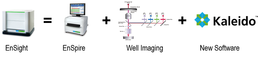
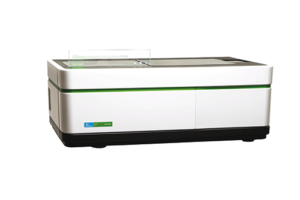
Multilabel Plate Readers
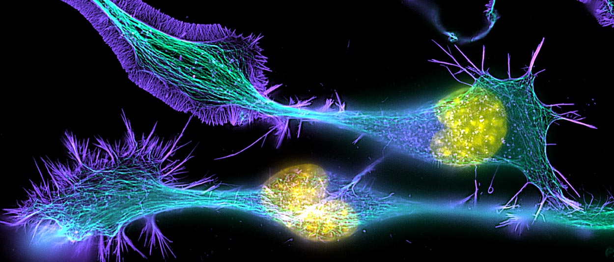
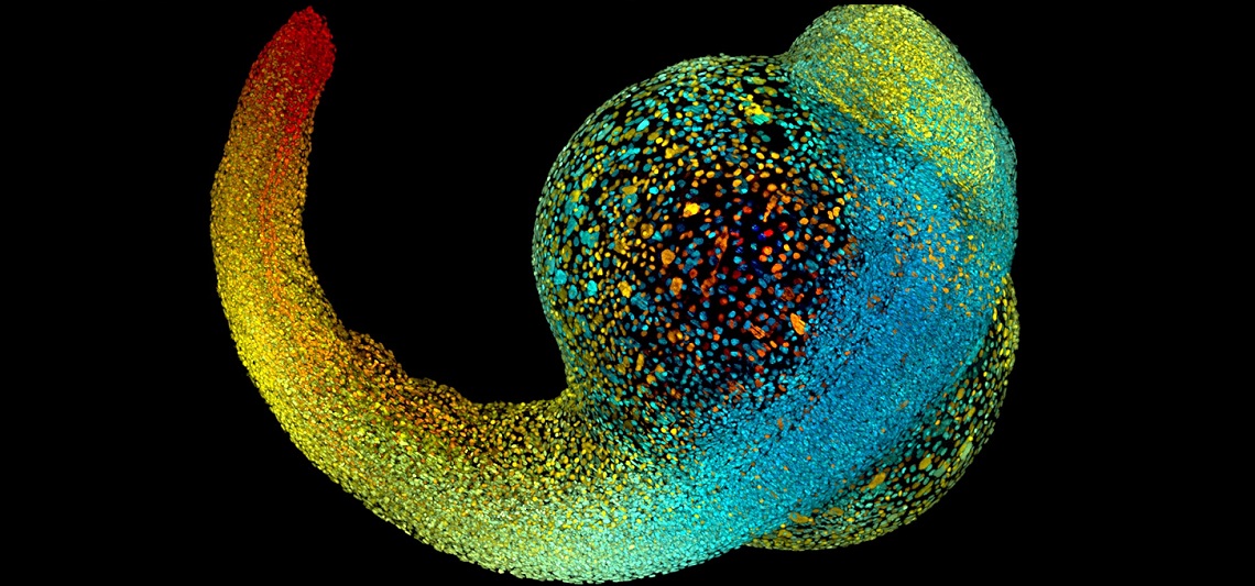
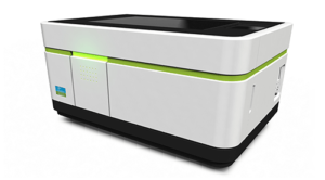
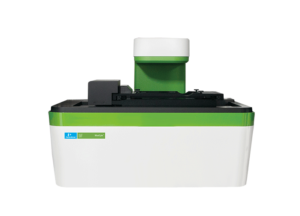
High Content Screening Systems
PerkinElmer High Content Screening allows scientists to image more samples and analyse more parameters so that they can answer complex biological questions. The complete analysis capabilities, makes the systems suitable for a wide range of applications from basic research to assay development and screening.
Opera Phenix
Operetta CLS
MuviCyte Live-Cell Imaging Kit
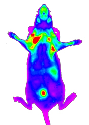
In Vivo Imaging & Analysis
ANTISEL through PerkinElmer offers a complete solution toolset for in vivo imaging. Our pre-clinical systems include 2D and 3D Fluorescence, Luminescence, Micro CT and PET imaging technologies as well as the industry’s widest selection of imaging reagents.
https://youtu.be/HGFPbM-9_JE